The human body under the microscope? See here some facts and interesting pictures of it.
Have you ever questioned what occurs beneath your skin’s surface and what is invisible to the human eye? Get ready for an incredible voyage as we explore the little gems that make up the complex fabric of the human body.
Our bodies under the microscope reveal a fascinating scene, from the busy cities of cells to the complex highways of blood vessels. And trust us when we say it’s jaw-dropping!
Come along as we get smaller and see the amazing ballet of microbes that keeps us going. If you haven’t read anything like this before, then you’re definitely in the right place. Here at Science in the World, we are looking to discover every little detail about the latest scientific discoveries that make the world spin.
Without further ado, let’s start the article and see some of the best pictures we could find of the human body cells under the microscope. And if you like it, don’t forget to hit that subscribe button.

1. Empty fat cells
Have you ever wondered how a fat cell looks under the microscope? Among the biggest cells in the human body are fat cells. Under the skin, they provide a thick layer of insulation that both cushions and stores energy.
The typical lipid (fat) deposits on the cells, which comprise most of the picture, have been eliminated to show the cell membranes’ honeycomb structure. The cells enlarge with more fat when we gain weight, and ultimately, new cells are added as well.
2. White blood cells
We are moving forward with the second thing that looks pretty interesting under the microscope, which is the white blood cells. These cells, sometimes referred to as leukocytes, come in a variety of forms, including neutrophils, lymphocytes, and monocytes. They play a huge role in the immune system.
White blood cells, despite their name, are colorless, but when seen under a microscope and stained with dye, they can take on a faint purple-to-pink hue. These little cells are round and include a unique core membrane known as the nucleus.
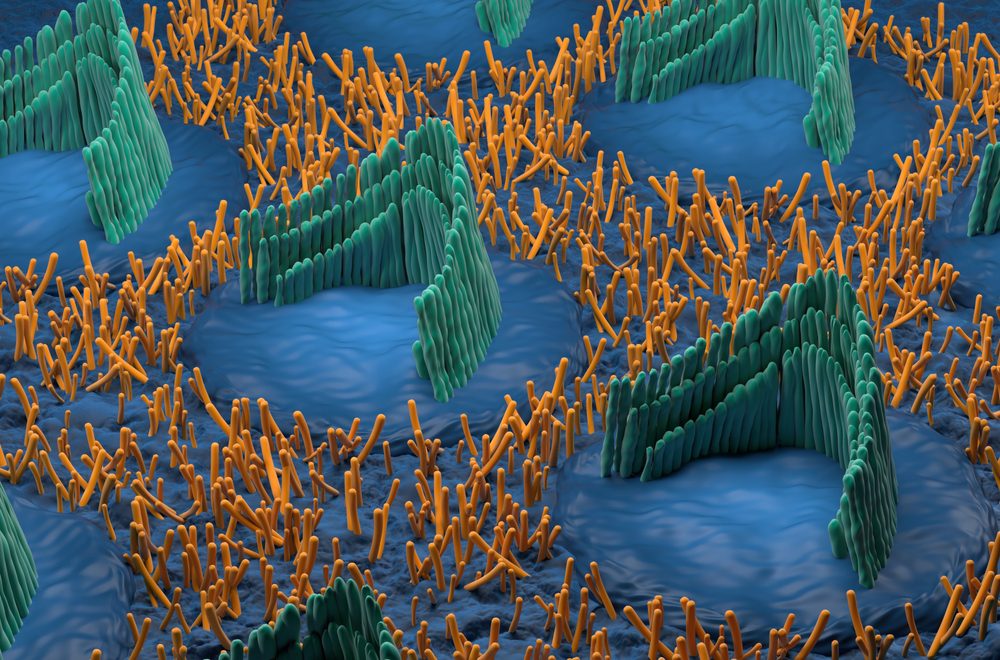
3. Human skin
The epidermis, the skin’s outermost layer, is made up of dead cells that are continually shed from underneath and replaced. High concentrations of the yellow protein keratin, which gives skin strength and waterproofness to protect internal organs, are found in these densely packed cells. Hair follicles—those small black ones—can also be seen when examining human skin under a microscope.
Does this article about the human body under the microscope make you curious about more scientific stuff? We got you! Check out You Under the Microscope 1st edition by Charlie Hatton. A smart and funny book about our human body. It can be found on Amazon for just $18.95 for the paperback format.
4. Tumor blood vessels
A tumor blood vessel looks chaotic and erratic under a microscope, with strange branching and differing sizes. Its patterns are twisted and disorganized, unlike typical vessels.
There is a noticeable increase in vascular density, which indicates the tumor’s need for nutrition. These abnormalities encourage the fast growth and spread of cancer, which adds to its severe character.
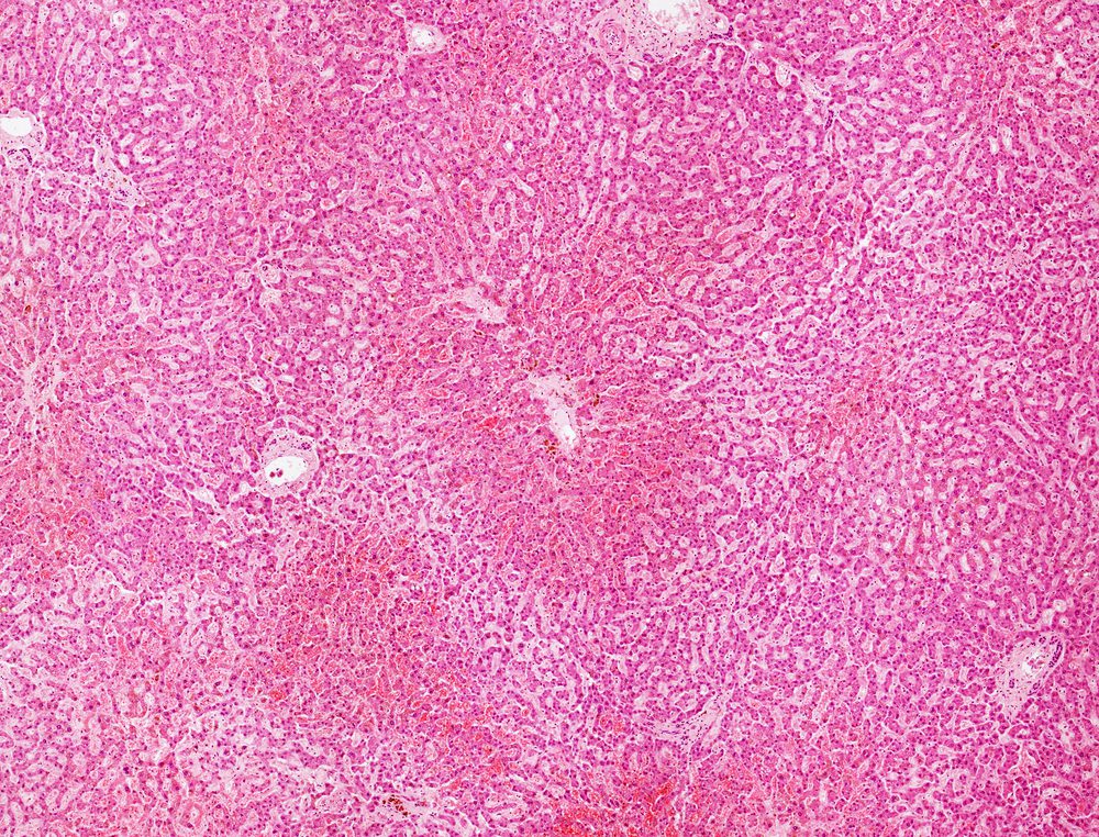
5. Liver Cells
Pretty amazing, right? For as long as your liver is healthy, you won’t really notice. Your liver is one of the biggest and most vital organs in your body, even though it functions “behind the scenes.”
It assists certain other organs in carrying out their duties and carries out more than 300 different tasks. This close-up shot of the liver under the microscope illustrates what a healthy liver looks like.
6. Influenza virus particles
Humans, pigs, birds, and horses can all contract influenza A viruses. The 2009 swine flu outbreak was brought on by the H1N1 strain. Each virus has a protective protein shell (the nucleocapsid, yellow) around its genetic imprint (the ribonucleic acid, pink).
The two types of proteins found in the enclosing lipid envelope (green) are called neuraminidase and haemagglutinin (the “H” and “N” in the strain designation), and their relative amounts identify the type of virus.
Flu viruses A and B are nearly identical when seen under the microscope. They have a spherical or filamentous form; the filamentous forms are frequently longer than 300 nm, while the spherical forms have a diameter of about 100 nm.
7. Bacteriophage
This bacteriophage resembles a bug or an ant at first glance. However, the virus is minuscule and has recently bonded to an E. coli bacteria (seen in blue under the microscope).
Phages, or bacteriophages, are viruses that exclusively reproduce and infect bacterial cells. They are acknowledged as the most prevalent biological agents on Earth and are found everywhere in the environment. Their size, appearance, and genetic structure exhibit remarkable diversity.
If this news didn’t make your skin crawl, then sit tight because we have a couple of more interesting things for you!
8. Gallstone crystals
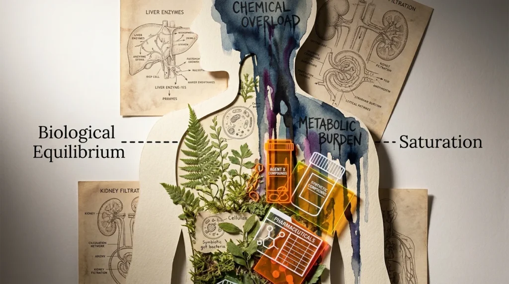
The primary component of gallstones is cholesterol, although they can also include calcium and bilirubin, which is a byproduct of aged red blood cells. When the bile’s chemical makeup is out of equilibrium, it develops in the gallbladder, which is where bile is expelled into the small intestine.
Gallstones often don’t cause any symptoms until they block the bile duct. Then they result in infection, jaundice, and excruciating agony. Under the microscope, these gallstones look pretty nice, a light violet color with blue hints. They’re also pretty spiky, too.
Are you looking to make your kids or grandkids interested in this scientific topic and be more into biology at school? Then you’ll like this particular book we recommend you to check out.
Explore, Experiment, and Discover the World of Science is an activity book recommended for kids over the age of 5 and it can be a great choice if you want to spend a fun but also interesting activity with your grandkids. Fun is guaranteed for both sides. It’s only $7.99 on Amazon at the moment of speaking.
9. Balancing stone from the inner ear
An otolith, which means “ear-stone” in old Greek, is a microscopic stone found in each ear that is responsible for our sense of balance. As may be seen on the surface of an otolith, deposits of calcium carbonate crystals cause the stones to accumulate in the inner ear.
These stones can occasionally go into the semicircular canal, a region of the inner ear. Your semicircular canal’s stones move in different ways depending on how you move your head. Fascinating, right?
10. Blood clot
Blood clots can form inside blood vessels or on the skin’s surface as a result of trauma. Overproduction of platelets might be the source of these internal clots, also known as “thrombi.” They might trigger a heart attack.
Under the microscope, they look like tiny squashed oranges trapped in a bold and tangled spider web.
Are you interested in reading more about this topic? Or do you have other suggestions for us? Don’t hesitate to tell us in the comments section and we will do our best to make your wishes come true.
Hey you! Are you still curious about other scientific facts? Before closing the page don’t forget to check out another favorite of ours How Long Can Humans Truly Live? 7 Fascinating Facts You Didn’t Know About The Human Life.